Cemental Dysplasia / Osseous Dysplasia Periapical Springerlink : People with fcod develop lesions in the jaw, were spots of normal bone are replaced with a mix of connective tissue and abnormal bone.
Cemental Dysplasia / Osseous Dysplasia Periapical Springerlink : People with fcod develop lesions in the jaw, were spots of normal bone are replaced with a mix of connective tissue and abnormal bone.. A benign, asymptomatic condition affecting the development of the periapical tissues. Periapical cemental dysplasia is a rare finding in the normal population. Tooth is normal to palpation, percussion and probings. It has a variable radiographic appearance depending on the phase at which it is diagnosed. Three types have been described:
Tooth is normal to palpation, percussion and probings. Found most commonly at the apex of mandibular anterior teeth in middle aged, black females. The vitality of the teeth was measured by two different testing methods. Kawai t, hiranuma h, kishino m, jikko a, sakuda m. It can be observed in the periapical area of vital teeth or in extraction sites.
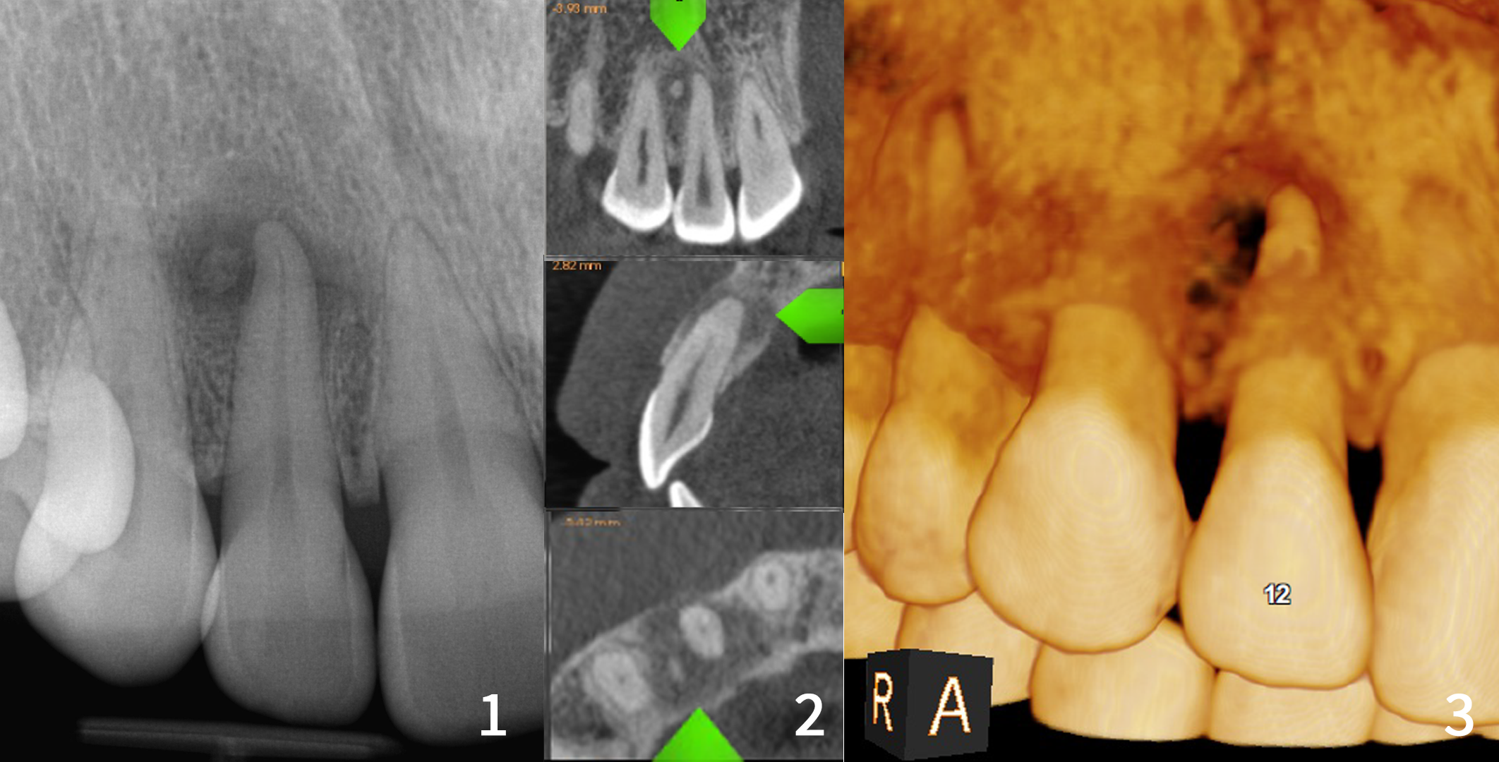
File Xray And Cbct Of Periapical Cementral Dysplasia Png Wikimedia Commons from upload.wikimedia.org Periapical cemental dysplasia is common in women with nf1. The correct diagnosis of pathological lesions of endodontic origin should allow for differentiation from those arising from other sources. Periapical cemental dysplasia is a rare finding in the normal population. Oral surg oral med oral pathol oral radiol endod 1999; Islam mn, cohen dm, kanter kg, stewart cm, katz j, bhattacharyya i. Three types have been described: Cbct shows the radiographic findings from. She was asymptomatic, with a prior rct on #22.
The vitality of the teeth was measured by two different testing methods.
The vitality of the teeth was measured by two different testing methods. She was asymptomatic, with a prior rct on #22. (b) pacd opacifies forming aradiographic ''target lesion'' in later stages of the disease. A total of 55 patients with nf1, 29 female and 26 male patients, were evaluated with orthopantomograms, supplemented with periapical radiographs if necessary. 3 cases of periapical cemental dysplasia with multiple lesions in both maxilla and mandible are reported. Islam mn, cohen dm, kanter kg, stewart cm, katz j, bhattacharyya i. Periapical cemental dysplasia in an endodontically treated tooth. Periapical cemental dysplasia is a rare finding in the normal population. It can be observed in the periapical area of vital teeth or in extraction sites. Smith s, patel k, hoskinson ae. The vitality of the teeth was measured by two different testing methods. A 42 yr old, white, female presented for evaluation of #22. The lesion occurs in and near the periodontal ligament around the apex of a tooth, usually a mandibular incisior.
Periapical cemental dysplasia is a benign condition mostly seen in patients over 20 years of age and is more common in women. A 42 yr old, white, female presented for evaluation of #22. The lesion occurs in and near the periodontal ligament around the apex of a tooth, usually a mandibular incisior. Periapical cemental dysplasia is a rare finding in the normal population. People with fcod develop lesions in the jaw, were spots of normal bone are replaced with a mix of connective tissue and abnormal bone.
Periapical Cemento Osseous Dysplasia Clinicopathological Features Anticancer Research from ar.iiarjournals.org She was asymptomatic, with a prior rct on #22. A 42 yr old, white, female presented for evaluation of #22. It can be observed in the periapical area of vital teeth or in extraction sites. Periapical cemental dysplasia is a rare finding in the normal population. The lesion occurs in and near the periodontal ligament around the apex of a tooth, usually a mandibular incisior. The correct diagnosis of pathological lesions of endodontic origin should allow for differentiation from those arising from other sources. 3 cases of periapical cemental dysplasia with multiple lesions in both maxilla and mandible are reported. Cbct shows the radiographic findings from.
A large nodular, irregular, radiopacity found within a large radiolucent area on the periapical radiograph.
A benign, asymptomatic condition affecting the development of the periapical tissues. People with fcod develop lesions in the jaw, were spots of normal bone are replaced with a mix of connective tissue and abnormal bone. It has a variable radiographic appearance depending on the phase at which it is diagnosed. Islam mn, cohen dm, kanter kg, stewart cm, katz j, bhattacharyya i. Kawai t, hiranuma h, kishino m, jikko a, sakuda m. (a) multiple confluent opacities in all four jaw quadrants. Periapical cemental dysplasia is a benign condition mostly seen in patients over 20 years of age and is more common in women. A total of 55 patients with nf1, 29 female and 26 male patients, were evaluated with orthopantomograms, supplemented with periapical radiographs if necessary. A 42 yr old, white, female presented for evaluation of #22. Its etiology is not fully understood, but possibly it is related to an unusual bone and cementum response to some local factor. Following surgery, biopsy material was submitted and diagnosed as periapical cemental dysplasia (pcd). It can be observed in the periapical area of vital teeth or in extraction sites. Periapical cemental dysplasia in an endodontically treated tooth.
A total of 55 patients with nf1, 29 female and 26 male patients, were evaluated with orthopantomograms, supplemented with periapical radiographs if necessary. Oral surg oral med oral pathol oral radiol endod 1999; It has a variable radiographic appearance depending on the phase at which it is diagnosed. Well circumscribed round, periapical lucencies usually of anterior mandibular teeth. 3 cases of periapical cemental dysplasia with multiple lesions in both maxilla and mandible are reported.
An Unusual Case Of Focal Cemento Osseous Dysplasia Occupying Nearly The Entire Maxillary Sinus And Arising From Adjacent Tissue Of Fused Teeth Sciencedirect from ars.els-cdn.com Epub 2007 apr 21 doi: Lesions are usually multiple, most frequently involve vital mandibular anterior teeth, surround the root apices, and are initially radiolucent (becoming more opaque as they mature). Following surgery, biopsy material was submitted and diagnosed as periapical cemental dysplasia (pcd). (a) multiple confluent opacities in all four jaw quadrants. 3 cases of periapical cemental dysplasia with multiple lesions in both maxilla and mandible are reported. A case of periapical cemental dysplasia (cementoma) is. The vitality of the teeth was measured by two different testing methods. It has a variable radiographic appearance depending on the phase at which it is diagnosed.
Oral surg oral med oral pathol oral radiol endod 1999;
Its etiology is not fully understood, but possibly it is related to an unusual bone and cementum response to some local factor. It can be observed in the periapical area of vital teeth or in extraction sites. A total of 55 patients with nf1, 29 female and 26 male patients, were evaluated with orthopantomograms, supplemented with periapical radiographs if necessary. Oral surg oral med oral pathol oral radiol endod 1999; Well circumscribed round, periapical lucencies usually of anterior mandibular teeth. Periapical cemental dysplasia is a benign condition mostly seen in patients over 20 years of age and is more common in women. The vitality of the teeth was measured by two different testing methods. Kawai t, hiranuma h, kishino m, jikko a, sakuda m. A case of periapical cemental dysplasia (cementoma) is. Periapical cemental dysplasia is a rare finding in the normal population. Periapical cemental dysplasia is common in women with nf1. Found most commonly at the apex of mandibular anterior teeth in middle aged, black females. Cbct shows the radiographic findings from.


0 Komentar